Carotid Angiography
Perhaps the most feared cardiovascular complication other than heart attack is stroke. Orlando Heart & Vascular Institute cardiologists are careful to evaluate all of our patients for risk of stroke as well as risk of heart attack. One important disease that can result is stroke is carotid artery obstruction. Just as atherosclerotic plaque can build up in the coronary arteries, similar plaque can build up in the carotid arteries which carry blood from the aorta to the brain. Severe blockage in the carotid arteries can eventually lead to stroke, even before the artery becomes completely blocked. In addition to physical exam when your cardiologist listens to your carotid arteries for a “bruit” with a stethoscope, non-invasive studies may be obtained including carotid ultrasonography, CT angiography (CTA) or MR angiography (MRA). While helpful, none of these tests is 100% accurate. If you are suspected of having severe carotid narrowing, your cardiologist may well recommend a carotid angiogram.


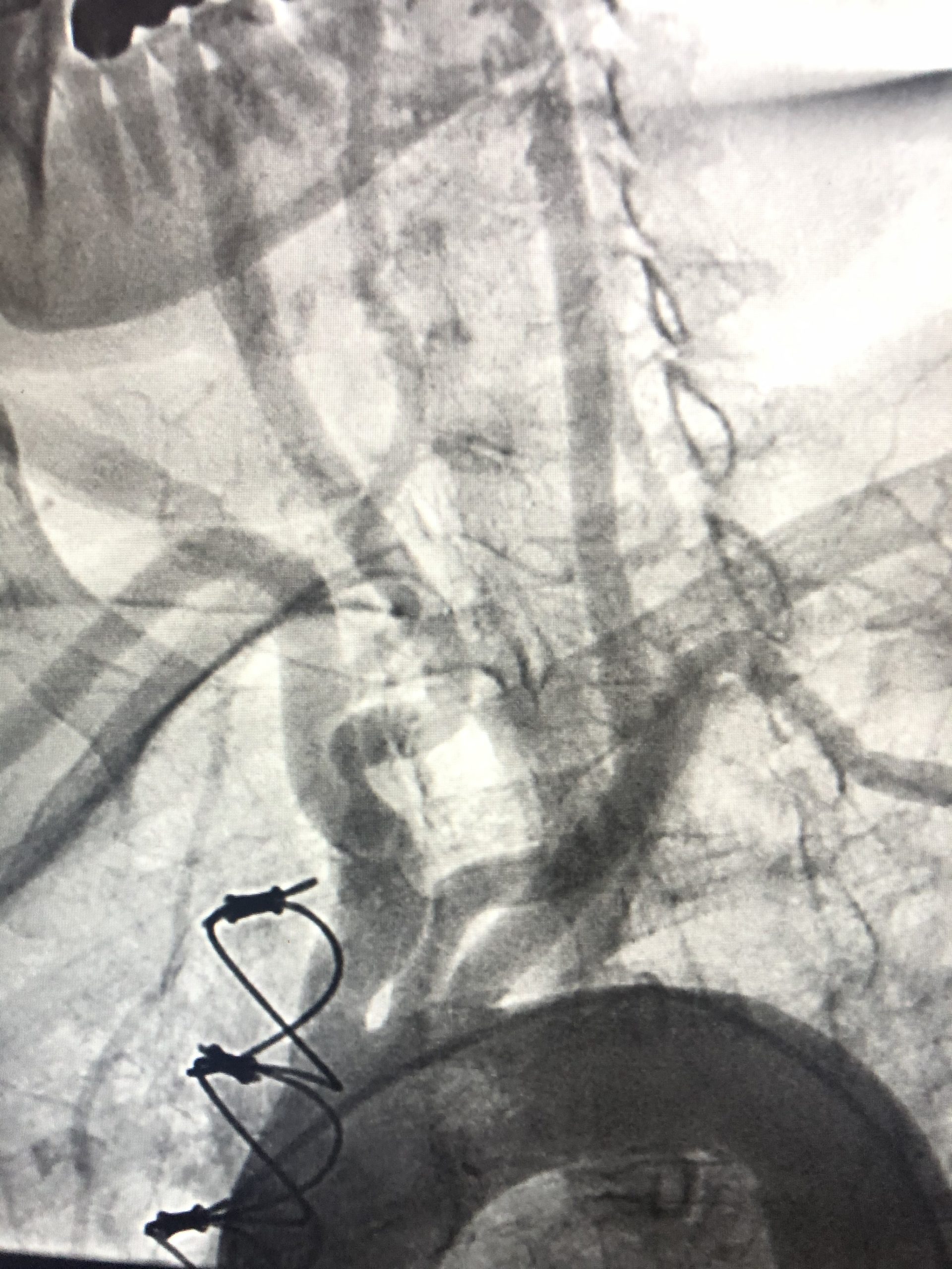
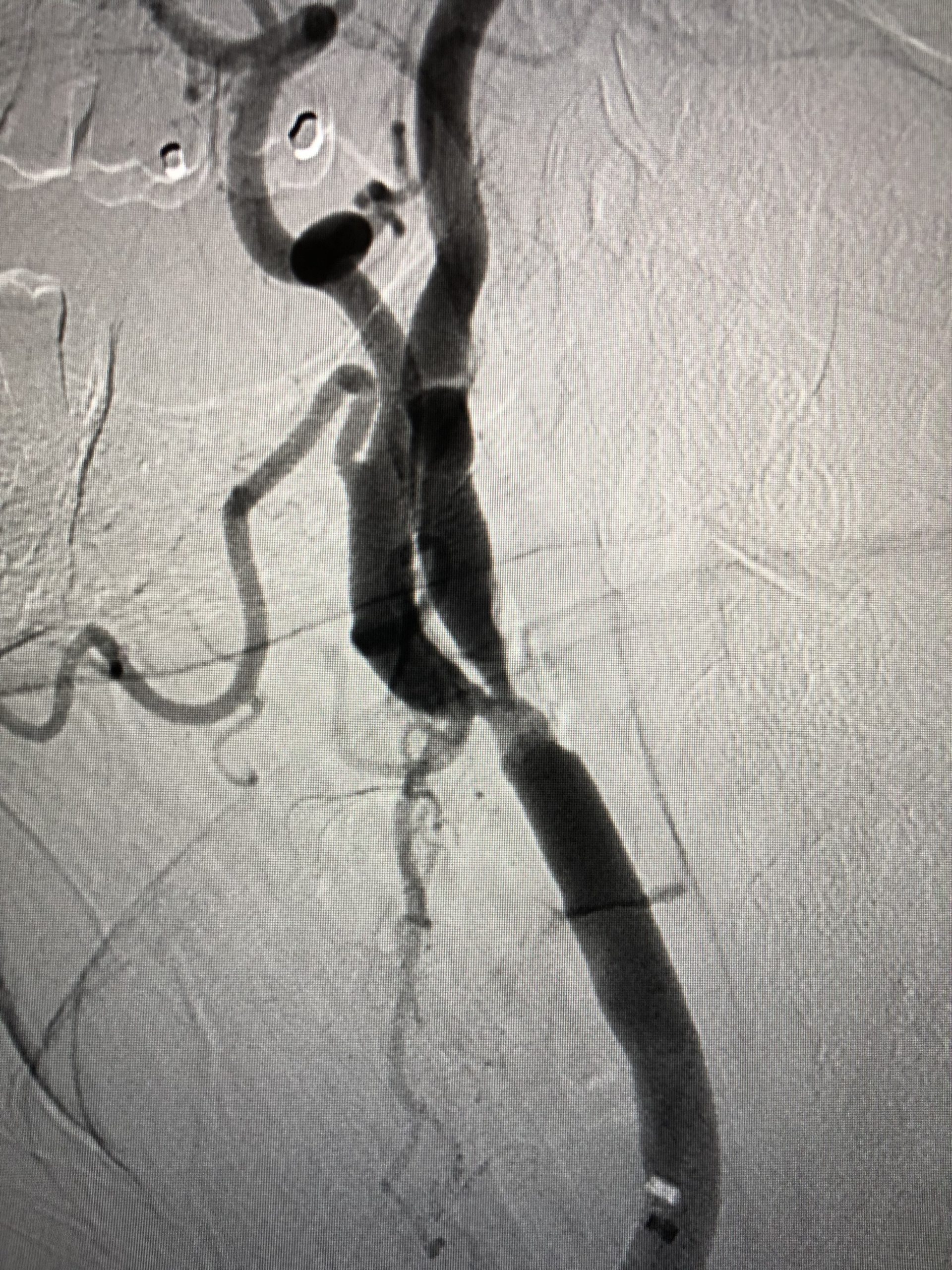
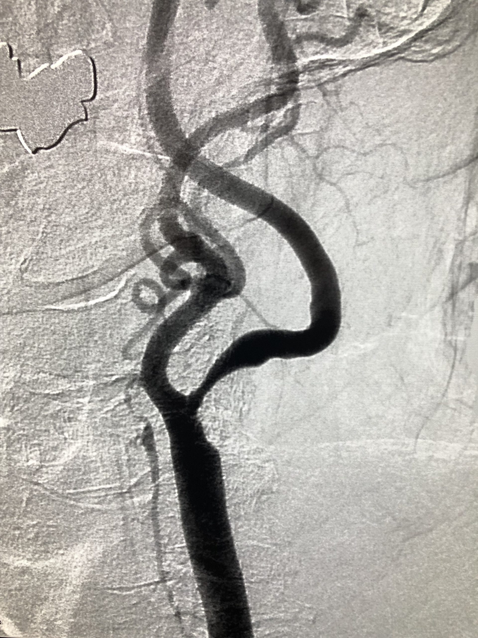
Similar to a heart catheterization, a carotid angiogram is obtained by advancing a catheter from the femoral artery in the groin up to the carotid arteries in the neck. Contrast can be injected and an angiogram obtained to evaluate the blockage with 100% accuracy. The blood vessels in the brain can be studied, as well. The carotid angiogram is often used to determine if a patient needs carotid revascularization, and whether carotid stenting or carotid surgery (“endarterectomy”) is the best option. Factors such as vessel tortuosity, calcification, and configuration of the aortic arch all contribute to determining the best treatment option for each patient.
Carotid angiography is a safe procedure with a low risk of complications (including a low risk of stroke) which can be performed in the hospital (with simultaneous stenting if indicated) or in the outpatient cath lab for diagnostic purposes only.


